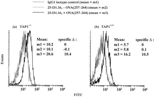
Figure 3.
Analysis of OVA(257–264):Kb complexes formed on viable TAP1−/− and TAP1+/+ macrophages by flow cytometry. Viable macrophages were pulsed with or without 5 μm OVA(257–264) at 37° for 2 hr. The cells were then stained with 25-D1.16 or isotype-matched control antibody. (a) TAP1−/− macrophages. (b) TAP1+/+ macrophages.

