
Figure 1.

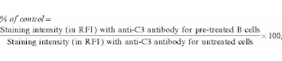
The effect of CR1 blockade on C3 fragment deposition on normal B cells, following in vitro AP activation. The reactivity, with rabbit–anti-huC3c or anti-C3d Ab, of C3 fragments deposited on B cells preincubated with the blocking mAb, 3D9, are expressed as a percentage of the reactivity of fragments on untreated cells, following 10 or 30 min of AP activation.*The vertical bars denote the 95% confidence intervals. *The murine IgG1 anti-huCR1 mAb, 3D9,7 was cleaved with pepsin to yield F(ab′)2 fragments, for use in the blockade of CR1 function, and FITC-conjugated IgG preparations of rabbit–anti-huC3c and anti-huC3d Ab (DAKO, Denmark) were digested with papain to generate their Fab fragments. PBL or Raji cells (ca. 1 × 10⁶ were preincubated with or without the 3D9 F(ab′)2 fragments (1 μg/ml) in phosphate-buffered saline/bovine serum albumin (PBS/BSA; 400 μl) for 1 hr at 4°. AP activation was performed for 10 or 30 min as described in Table 1. The cells were then washed and incubated with saturating concentrations of polyclonal FITC-anti-C3c or -anti-C3d, R-PE anti-huCD19 and huIgG in PBS/BSA for 2 hr on ice. Flow cytometric analysis was performed essentially as described in Table 1, although in this case the data was expressed as:
where RFI = relative fluorescence intensity units.
 |
1 |