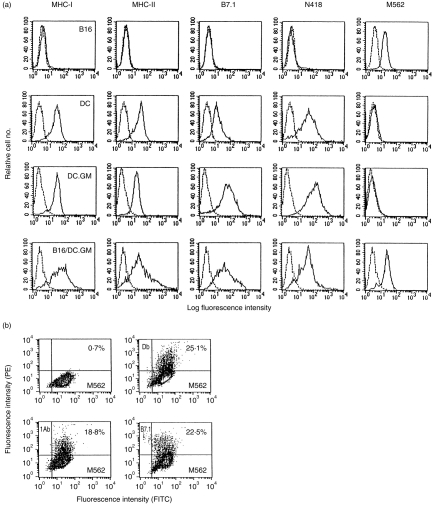
Figure 1.
Phenotypic analysis of B16, granulocyte–macrophage colony-stimulating factor gene-modified dendritic cells (DC.GM) and their fusion hybrid. (a) B16, DCs, DC.GM and purified B16/DC.GM fused cells were analysed by flow cytometry for expression of major histocompatibility complex (MHC) class I (Db, Kb) and class II (IAb) antigens, co-stimulatory molecule (B7.1), DC marker CD11c (N418) and B16 tumour marker (M562), as detailed in the Materials and methods. (b) DC.GM were fused with B16 at a fusion ratio of 10:1 by polyethylene glycol (PEG). A bulk B16/DC.GM fusion preparation was cultured overnight and the non-adherent cells (unfused DCs and dead cells) were removed. The retained adherent cells were harvested and double stained with M562 monoclonal antibody (mAb), or with M562 plus mAbs against IAb, or Db or B7.1. Numbers in the quadrants of dual-parameter histograms represent the percentage of cells contained within that quadrant. FITC, fluorescein isothiocyanate; PE, phycoerythrin.

