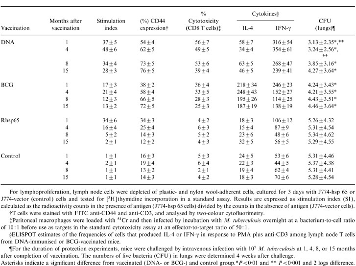
Table 5.
Correlation between the duration of protection against tuberculosis challenge infection and development of immunological memory and cytokine production at several intervals after vaccination

For lymphoproliferation, lymph node cells were depleted of plastic- and nylon woll-adherent cells, cultured for 3 day with J774-hsp 65 or J774-vector (control) cells and tested for [3H]thymidine incorporation in a standard assay. Results are expressed as stimulation index (SI), calculated as the radioactivity counts in the presence of antigen (J774-hsp 65 cells) divided by the counts in the absence of antigen (J774-vector cells).
†T cells were stained with FITC anti-CD44 and anti-CD3, and analysed by two-colour cytofluorimetry.
‡Peritoneal macrophages were loaded with 51Cr and then infected by incubation with M. tuberculosis overnight at a bacterium-to-cell ratio of 10: 1 before use as targets in the standard cytotoxicity assay at an effector-to-target ratio of 50: 1.
§ELISPOT estimates of the frequencies of cells that produced IL-4 or IFN-γ in response to PMA plus anti-CD3 among lymph node T cells from DNA-immunised or BCG-vaccinated mice.
¶For the duration of protection experiments, mice were challenged by intravenous infection with 105 M. tuberculosis at 1, 4, 8, or 15 months after completion of vaccination. The numbers of live bacteria (CFU) in lungs were determined 4 weeks after challenge.
Asterisks indicate a significant difference from vaccinated (DNA- or BCG-) and control grroup. *P < 0·01 and **P < 0·001 and 2 logs difference.
