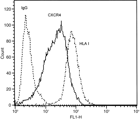
Figure 2.
Flow cytometry histograms of CXC4 on A549 epithelial cells. Cells were stained with either anti-CXCR4 or anti-HLA class I antibody followed by FITC-conjugated anti-IgG. Controls received equivalent concentrations of isotype-matched IgG. For all panels, data are shown as cell number vs. the relative fluorescence. Each histogram shows data from a single representative experiment although each analysis was repeated at least three times. A549 cells abundantly express CXCR4 at the cell surface with almost comparable levels to HLA class I antigen.

