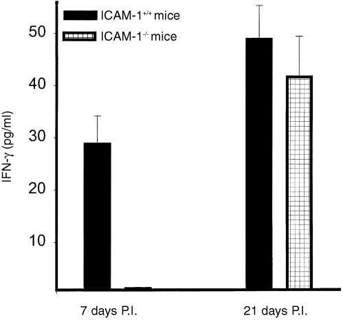
Figure 2.
Recruitment of chlamydia‐reactive Th1 cells after chlamydial genital infection of ICAM‐1–/– and ICAM‐1+/+ mice. Nylon wool‐purified T cells were isolated at the indicated time‐points from genital tract tissues of infected female ICAM‐l–/– and ICAM‐1+/+ mice. The T cells were stimulated with APCs and chlamydial antigen for 5 days and the amounts of IFN‐γ in the culture supernatants were measured by ELISA as described in the Materials and Methods section. The concentrations of IFN‐γ are expressed as the mean (pg/ml) of results from different experiments. Control cultures that contained T cells and APCs but without chlamydial antigen did not show any measurable amounts of IFN‐γ and so the data are not presented in the results shown.

