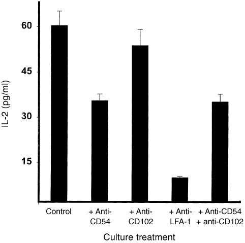
Figure 6.
Contribution of LFA‐1 and its ligands to immune T‐cell activation. Nylon wool‐purified T cells were isolated from the spleens of genitally infected ICAM‐1+/+ mice. The T cells (2 × 10⁵/well) were stimulated with 10 µg/ml of chlamydial antigen, APCs from ICAM‐1+/+ mice, in the presence of neutralizing mAbs directed against LFA‐1, CD54 (ICAM‐1), CD102 (ICAM‐2) or a combination of anti‐CD54 and CD102. After 72 hr of incubation, the amounts of IL‐2 in the culture supernatants were measured by ELISA as described in the Materials and Methods section. The concentrations of IL‐2 are expressed as the mean (pg/ml) of results from different experiments. Cultures containing T cells and APCs but without chlamydial antigen did not show any measurable amounts of IL‐2 and so the data are not presented in the results shown.

