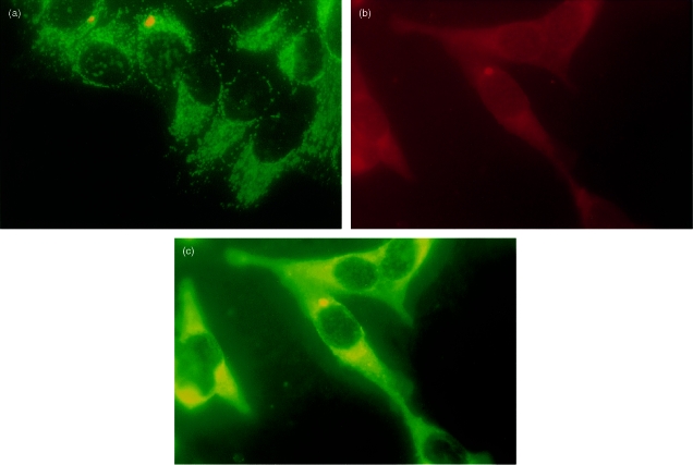
Figure 2.
Double fluorescence staining on Hep‐2 cells. (a) Monoclonal antibody (mAb) B‐33 (orange staining), anti‐human lysosome associated membrane glycoprotein‐1 (anti‐hLAMP‐1) and hLAMP‐2 antibody (green staining). (b) mAb B‐33 (red staining). (c) Anti‐cation‐independent mannose 6‐phosphate receptor (anti‐CI‐MPR) serum (green staining).

