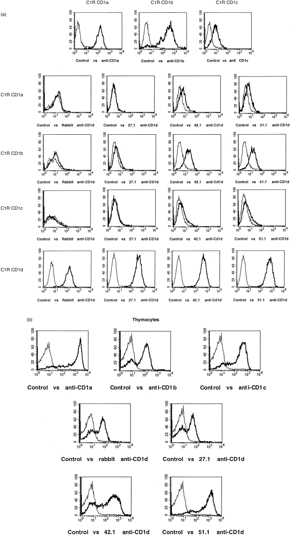
Figure 1.
Specificity of anti-CD1d antibodies and thymocyte CD1d expression by indirect immunofluorescence. (a) Polyclonal rabbit anti-CD1d and mouse monoclonal 27.1, 42.1 and 51.1 anti-CD1d antibodies were tested by indirect immunofluorescence against C1R cells stably transfected with CD1a, b, c or d, as indicated. The top three panels (C1R CD1a, C1R CD1b and C1R CD1c) are the CD1a, b and c transfectants stained with anti-CD1a (OKT6), anti-CD1b (4A76) and anti-CD1c (M241) monoclonal antibodies (mAbs), respectively. The thick lines represent specific antibodies and the thin lines are control antibodies (normal rabbit or mouse immunoglobulin G [IgG]). (b) Freshly isolated human thymocytes were stained by indirect immunofluorescence with the indicated specific (thick lines) or control (thin lines) antibodies.

