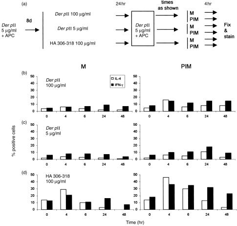
Figure 5.
Cytokine expression in response to antigen re‐stimulation in cells recently tolerized or stimulated. Cloned AC1.1 cells were treated as outlined in (a). Resting cells were incubated for 24 hr with (b) specific antigen Der p II peptide (28–40) at 100 μg/ml; (c) Der p II peptide (28–40) at 5 μg/ml, or (d) irrelevant peptide HA (306–318) at 100 μg/ml. After extensive washing, all three groups were stimulated for various time‐intervals with Der p II at 5 μg/ml (second antigenic stimulus), then incubated for 4 hr with either PI and monensin (PIM) or monensin alone (M). Histograms show percentages of cells positive for IFN‐γ and IL‐4, based on matched isotype controls.

