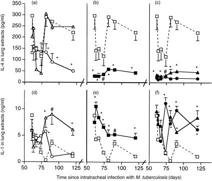
Figure 4.
ELISA anaysis of IL-4 (a–c) and IL-1 (d–f) in the lungs of BALB/c mice infected with M. tuberculosis H37Rv, and treated with the indicated regimen from day 60. The higher dose of M. vaccae or AED caused decreased IL-4 content, in agreement with the immunohistochemical data. There was also increased expression of IL-1, particularly in the combined therapy group. (□) control infected mice; (▵) mice treated day 60 and day 90 with M. vaccae 0·1 mg; (○) mice treated day 60 and day 90 with M. vaccae 1·0 mg; (▪) mice treated from day 60, three times per week with AED + corticosterone; (▴) AED + corticosterone and 0·01 mg M. vaccae; (•) AED + corticosterone and 1·0 mg M. vaccae. Error bars are SD. *P < 0·005, #P < 0·025, relative to controls. The trough on days 63, 67 and 74 is unexplained.

