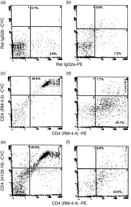
Figure 1.
CD4 protein on vaginal lymphoid cells by flow cytometry. Lymph node cell (106) and collagenase-digested vaginal lymphoid-like cells (105) from CBA/J mice were labelled with biotinylated (plus cychrome [CYC]-conjugated streptavidin) RM-4.5 or H129.19 and phycoerythrin (PE)-conjugated RM-4.4 epitope-distinct anti-CD4 antibodies. RM-4.5 and H129.19 anti-CD4 antibodies recognize an epitope in domain 1 of the CD4 protein, whereas RM-4.4 antibodies recognize an epitope in domain 3. Flow cytometric dual staining of lymph node cells (a, c and e) or vaginal lymphoid cells (b, d and f) with RM-4.4 and RM-4.5 anti-CD4 antibodies (c and d) and RM-4.4 and H129.19 anti-CD4 antibodies (e and f) is shown together with isotype control antibodies (a,b). The percentage in each quadrant represents the fluorescent positive cells within the lymphoid-like cell limits. Data shown are a representative of two experiments using 10 mice per experiment.

