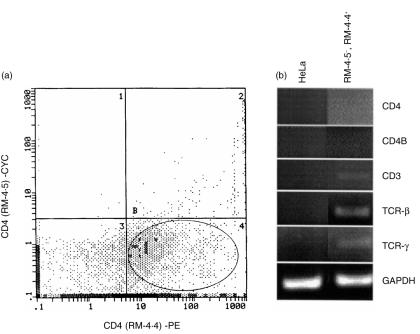
Figure 5.
lack of CD4 mRNA expression in RM-4.5– RM-4.4+ CD4+ vaginal cells. (a) Vaginal lymphoid cells extracted from CBA/J mice by collagenase-digestion and further purified by Ficoll–Paque® density gradient centrifugation were dual labelled with RM-4.4 and RM-4.5 anti-CD4 antibodies. Those cells that stained positively with RM-4.4 but not RM-4.5 monoclonal antibodies (RM-4.5– RM-4.4+) were FACS sorted. The RM-4.5– RM-4.4+ CD4+ cell-sorted population is represented in the encircled gate of quadrant B4. HeLa cells (5 × 10⁵) were added to the sorted cell population and pelleted. Total RNA was extracted from RM-4.5– RM-4.4+-plus HeLa cells or HeLa cells alone, reverse transcribed and subjected to PCR using a high-efficiency Taq DNA polymerase and primers designed to amplify CD3, CD4, CD4B, TCR β-, and TCR δ chain constant regions of murine systemic T lymphocytes (b). The CD4 and CD4B amplification products shown are from column-purified primary amplification products subjected to a second round of amplification. Amplification with primers for GAPDH was used as an internal control. The figure is representative of two experiments using 40 mice in each experiment.

