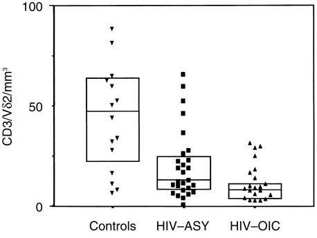
Figure 1.
γδ T-cell subset distribution in the peripheral blood of asymptomatic subjects infected with human immunodeficiency virus (HIV-ASY) and in HIV-infected patients with opportunistic infections/co-infections (HIV-OIC). γδ T-cell subset distribution was analysed ex vivo by flow cytometry in peripheral blood mononuclear cells (PBMC) from healthy donors (controls, n = 15) and HIV-positive subjects (n = 47: HIV-ASY, n = 28; HIV-OIC, n = 19). Vδ2 T-cell absolute numbers are shown. Lines represent group medians and boxes represent interquartile ranges. Kruskal–Wallis H-test results with Bonferroni correction: χ2 = 15·020, d.f. = 2, P = 0·005.

