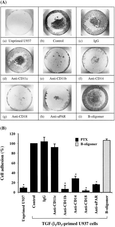
Figure 1.
Pertussis toxin (PTX)-induced myeloid cell adhesion in serum. (A) Representative photographs showing PTX-induced myeloid cell adhesion in a 96-well plate. Panel (a) represents unprimed U937 cells and panels (b) to (i) represent transforming growth factor-β1/1,25-(OH)2 vitamin D3 (TGF-β1/D3)-primed U937 cells. Panels (a) to (h) display the adherent responses of myeloid cells to PTX holotoxin (10 µg/ml) in the presence and absence of mouse monoclonal antibodies (mAbs) (2 µg/ml) directed against CD11c, CD11b, CD14, CD18 or urokinase receptor (uPAR). Panel (i) shows the adherent response of myeloid cells to PTX B-oligomer (3 µg/ml) alone. (B) Cumulative results showing the effects of different mAbs on PTX-induced myeloid cell adhesion. Each value represents the mean ± SEM of four to six experiments performed in duplicate and is expressed as the percentage of control adherent responses to PTX. *P < 0·05 compared with primed U937 cell controls. IgG, immunoglobulin G.

