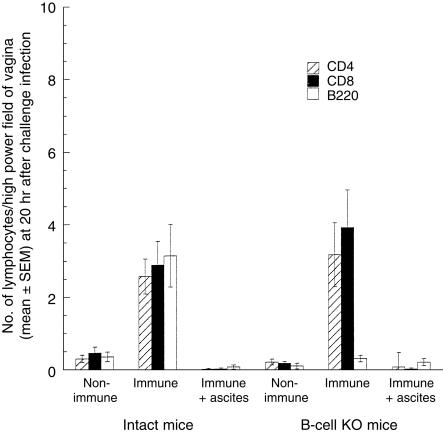
Figure 1.
Lymphocytes in vaginae of non-immune, immune and T-cell-depleted immune intact and B-cell knockout (KO) mice 20 hr after vaginal challenge with wild-type virus. The numbers of CD4+ and CD8+ cells in the vaginae of immune/challenged intact and B-cell KO mice were not significantly different (CD4, P = 0·93; CD8, P = 0·49). The small numbers of B cells observed in the B-cell KO groups are the result of non-specific staining, as staining with an isotype-matched control antibody in the intact immune group yielded a similar value of 0·2 cells per high power field. These counts arise because it is occasionally difficult to distinguish the membrane staining of lymphocytes from the endogenous fluorescence of granulocytes.

