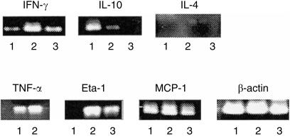
Figure 5.
RT-PCR analysis of the cytokine mRNA expression by the DN TCRαβ+ T cells after MCMV infection. C57BL/6 mice (18–20 mice per group) were infected intraperitoneally with 1 × 10⁵ PFU MCMV and plastic non-adherent PEC were harvested on days 0 and 5 after infection. The DN TCRαβ+ T cells were enriched by using dynabeads and MACS microbeads as described in the Materials and Methods. The CD4+ T cells were similarly enriched from the non-adherent PEC of the same mice on day 5 after MCMV infection and were used as a positive control. Total RNA from these purified cells was extracted and the cDNA was amplified by PCR using the specific cytokine sense and antisense primers. The sense and antisense primers of β-actin were used as control. The respective cytokine bands of lanes 1, 2 and 3 are for DN TCRαβ+ T cells on day 0, DN TCRαβ+ T cells on day 5 and CD4+ T cells on day 5 after MCMV infection, respectively. The figure represents one of two independent experiments with similar results.

