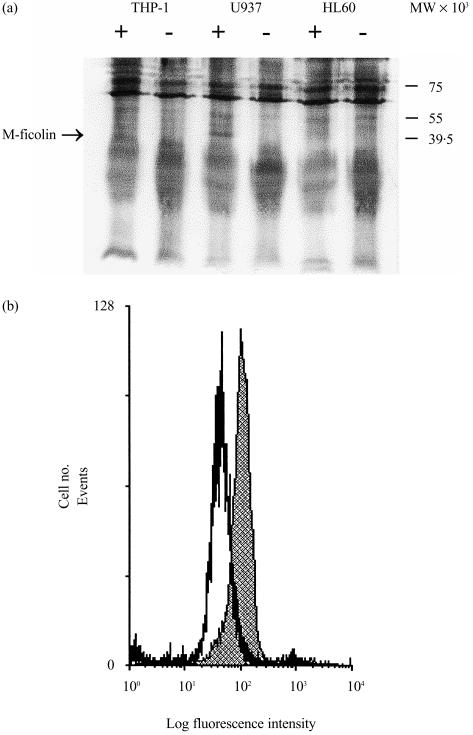
Figure 2.
Detection of M-ficolin on monocytoid cells. (a) Detection of M-ficolin on HL60, U937 and THP-1 cells by immunoprecipitation of biotinylated surface proteins. Surface-biotinylated cells were lysed using 0·5% (v/v) Nonidet P-40 (NP-40) and the supernatant precleared with Protein A–Sepharose. The supernatant was then incubated with either non-immune (−) or rabbit anti-fibrinogen-like (FBG) (+) rabbit serum and then with Protein A–Sepharose. The resin was harvested by centrifugation and, upon washing, eluted with sodium dodecyl sulphate–polyacrylamide gel electrophoresis (SDS–PAGE) sample buffer. The eluted proteins were separated by SDS–PAGE on a 12·5% (w/v) gel and electroblotted onto nitrocellulose membranes. Biotinylated cell surface proteins were detected with alkaline phosphatase-conjugated streptavidin and visualized using nitroblue tetrazolium (NBT) and 5-bromo-4-chloro-3-indolyl phosphate (BCIP). (b) Detection of M-ficolin on U937 cells by flow cytometry. U937 cells were washed and briefly surface cross-linked with sulpho-ethyleneglycobis(succinimidylsuccinate) (sulpho-EGS). The cells were blocked and then incubated with either non-immune or anti-FBG rabbit serum. After washing, the cells were further incubated with fluorescein isothiocyanate (FITC)-conjugated goat anti-rabbit F(ab′)2. After washing, the cells were fixed and analysed by flow cytometry on a Coulter Elite Epics ESP flow cytometer. The shaded profile denotes fluorescence detected on U937 cells with the anti-FBG antiserum while the open profile represents signals detected with non-immune rabbit serum.

