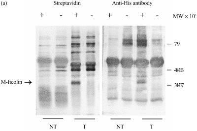
Figure 3.
Immunoprecipitation of biotinylated surface proteins of transfected COS-7 cells. Non-transfected COS-7 cells (NT), or those transfected with the His-tagged M-ficolin expression construct pcDNA3/FC (T), were surface biotinylated. The cells were lysed in 0·5% (v/v) Nonidet P-40 (NP-40) and the supernatants were, upon preclearance with Protein A–Sepharose, first incubated with either non-immune (−) or anti-FBG (+) rabbit serum and then with Protein A–Sepharose. Resin-bound proteins were, after washing, eluted with sodium dodecyl sulphate–polyacrylamide gel electrophoresis (SDS–PAGE) sample buffer and separated on a 12·5% (v/v) gel. The separated proteins were electroblotted and probed with either alkaline phosphatase-conjugated streptavidin or a mouse anti-His monoclonal antibody. The anti-His antibody was further probed with alkaline phosphatase-conjugated goat anti-mouse immunoglobulin G (IgG). The blots were developed with nitroblue tetrazolium (NBT) and 5-bromo-4-chloro-3-indolyl phosphate (BCIP).

