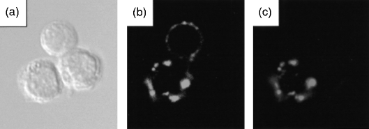
Figure 1.
Double labelling of B cells and their analysis using confocal microscopy. Cell suspensions from mouse mesenteric lymph node were stained with NeuAcα2,6Galβ1,4Glc coupled to biotinylated polyacrylamide (α2,6-PAA) followed by streptavidin-coupled fluorescein isothiocyanate (FITC), and then labelled with rat anti-mouse immunoglobulin D (IgD) followed by anti-rat-TRITC. (a) Phase contrast image of three lymph node cells. (b) Staining for IgD shows that two of the three cells are IgD+ B cells. (c) Staining with α2,6-PAA reveals that one of the three cells is a IgD+ B cell that is able to bind α2,6-PAA.

