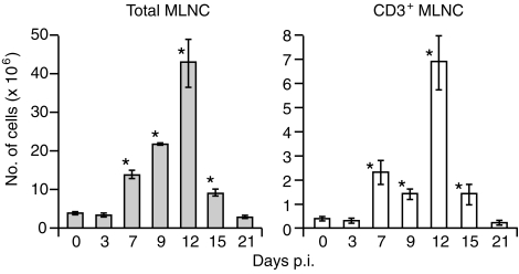
Figure 1.
Cellularity of mesenteric lymph nodes (MLNs) from T-cell receptor (TCR)-β–/– mice infected with Eimeria vermiformis. MLN cells were harvested, at the time-points indicated on the figure, from TCR-β–/– mice infected with 1000 sporulated oocysts. CD3+ cells were enumerated using flow cytometry with fluorescein isothiocyanate (FITC)-conjugated anti-CD3. Results are expressed as mean ± SE (n = 3 at each time-point). *Significant difference versus uninfected mice analysed in parallel (P < 0·05). p.i., postinfection.

