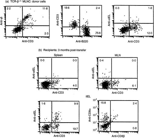
Figure 2.
Reconstitution of T-cell receptor (TCR)-(β × δ)–/– mice with mesenteric lymph node (MLN) cells from TCR-β–/– mice: phenotypic analysis. (a) TCR-β–/– MLN cells; donor cells. (b) Recipients, 3 months post-transfer. Representative flow cytometry plots from at least four individual mice are presented. Antibodies used were phycoerythrin (PE) -conjugated anti-γδ, -anti-CD3, -anti-B220, -anti-αIEL, and -anti-CD8-α, and fluorescein isothiocyanate-conjugated anti-CD3 and -anti-CD8β. iIEL, intestinal intraepithelial lymphocytes.

