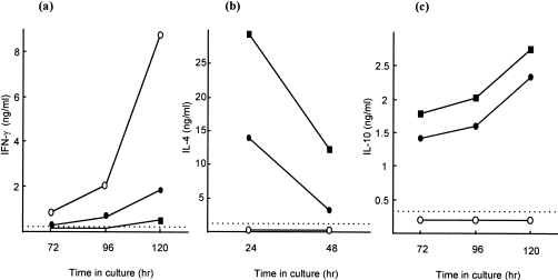
Figure 1.
Cytokine-secretion profiles of unfractionated, allergen-activated lymph node cells (LNC). Mice were exposed topically to 10% trimellitic anhydride (TMA) in acetone : olive oil (AOO) (•), 1% 2,4-dinitrochlorobenzene (DNCB) in AOO (○), or 0·5% fluorescein isothiocyanate (FITC) in dibutyl phthalate : acetone (DBP) (▪). Thirteen days after the initiation of exposure, draining auricular lymph nodes were excised and a single-cell suspension of LNC isolated. Supernatants were prepared after culture of LNC for different periods of time in the absence (interferon-γ [IFN-γ] and interleukin [IL]-10) or presence (IL-4) of 2 µg/ml of concanavalin A (Con A). IFN-γ (a), IL-4 (b) and IL-10 (c) concentrations were measured by using cytokine-specific enzyme-linked immunosorbent assay (ELISA). Results from a single representative experiment are presented. The limit of detection for each ELISA is indicated by the horizontal broken line.

