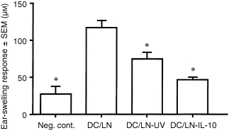
Figure 2.

CH‐inducing capacity of dendritic cells obtained from lymph nodes from IL‐10‐treated mice. Panels of mice received IL‐10 (200 ng) injections intradermally into abdominal skin. Eight hours later, lymph nodes not draining the injected skin (all except axillary and inguinal) were removed. Lymphoid cell suspensions were separated on density gradients using Percoll solution 1·075 g/dl, layered over 1·035 g/dl solution and centrifuged. Interface cells were collected and placed in Petri dishes at 37° for 60 min, after which non‐adherent cells were discarded. The adherent cells were then incubated at 37° overnight and non‐adherent cells were collected as cell suspension enriched for DCs. As positive control, cells were prepared from lymph nodes of untreated mice. DC‐enriched suspensions derivatized with DNFB in vitro were injected into footpads of naive syngeneic mice (2 × 10⁴ cells in 100 µl per mouse). Five days later CH was assayed as described in the legend to Fig. 1. Bars represent mean ear‐swelling responses ± SEM for groups of five mice each. Asterisks indicate mean responses significantly less than the positive control (P < 0·0005).
