Figure 1.

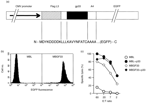
(a) The gp33 minimal epitope gene construct. The LCMV GP minimal epitope was flanked by three leucines and four alanines. For detection the FLAG tag was fused N-terminally and EGFP C-terminally to the minimal epitope. Expression of the fusion protein is driven by the cytomegalovirus (CMV) promoter. The expression plasmid was named pEGFPL33A. (b) Expression levels of the minimal epitope fusion protein. MBL-2 B-cell lymphoma cells were stably transfected with the fusion gene construct and expression levels of the minimal epitope gene product were determined by flow cytometry. One representative clone MBGF33 is shown. Non-transfected MBL-2 cells served as negative controls. (c) Specific lysis of MBGF33. Transfectants served as 51Cr-labelled target cells in a 5-hr cytotoxicity assay. LCMV-specific splenic effector CTL were obtained from mice which had been infected i.v. with 200 PFU of LCMV WE 8 days previously. To evaluate maximal antigen presentation and cytotoxicity the respective target cells were pulsed with gp33 peptide. Non-transfected MBL-2 cells served as controls. Spontaneous release was < 20% for all targets. A representative example of two independent experiments is shown.
