
Figure 5.
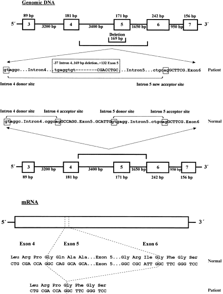
Schematic representation of the homozygous mutation in the leucocyte adhesion deficiency (LAD) patient. The 169-bp genomic deletion is depicted in the upper part of the figure. Exons are shown by thick boxes and introns by thick horizontal lines. Intron sequences are in lowercase letters and exon sequences in uppercase letters. Acceptor and donor splicing sites are also boxed and normal or pathological splicing is depicted by lines with small arrows. The 171 bp ‘in-frame’ mRNA deletion caused by the loss of the intron 4 acceptor splicing site and total exon 5 skipping, as well as the putative translated protein, are depicted in the lower part of the figure.