
Figure 2.
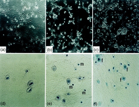
Morphology of fibroblasts and mast cells, co-cultured in three-dimensional lattices by phase contrast microscopy (a–c): (a) fibroblasts alone, (b) fibroblasts and mast cells at a 1 : 1 ratio, (c) fibroblasts and mast cells at a 1 : 5 ratio. Mast cells are indicated by arrows. Histopathological analysis of three-dimensional lattices 24 hr after the gel casting (d–f): H & E stain of fibroblasts alone (d), and fibroblasts and mast cell (m) co-cultures (1 : 1 ratio) (e), (f) toluidine blue stain of fibroblasts (f) and mast cell co-cultures (1 : 5 ratio). Magnifications: a–c, ×40; d, ×400; e,f, ×200.