
Figure 3.
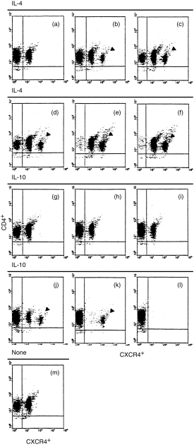
Double colour flow cytometric analysis of the kinetics of CXC chemokine receptor 4 (CXCR4) expression on human peripheral CD4+ T lymphocytes. The cells were stimulated either with IL-4 (10 ng/ml) for different time intervals indicated as (a, 1 hr), (b, 2 hr), (c, 4 hr), (d, 8 hr), (e, 16 hr) and (f, 24 hr), respectively, or with IL-10 (10 ng/ml) for different time intervals indicated as (g, 1 hr), (h, 2 hr), (I, 4 hr), (j, 8 hr), (k, 16 hr) and (l, 24 hr), respectively. (m) Freshly isolated unstimulated CD4+ T cells. The cells were then stained with anti-CXCR4 monoclonal antibody as described in the Materials and methods. The percentages of CXCR4+ cells are given in the Results. Arrows indicate the bright fraction of CXCR4+ cells. The data were taken from a single experiment, which was representative of each of three similar experiments performed.