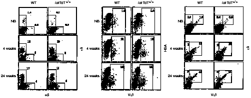
Figure 2.

Flow cytometric analysis of skin. Epidermal cells from wild-type (WT) and lck-promoter terminal deoxynucleotidyl transferase (lckTdT)+/+ mice at birth and at 4 and 24 weeks of age were stained for αβ/γδ, Vγ3/heat stable antigen (HSA) and Vγ3/γδ, and analysed by fluorescence-activated cell sorter (FACScan). All nucleated cells gated by forward scatter and side scatter were analysed. Numbers indicate the percentages of cells stained for a particular phenotype in the respective boxed regions. Representative data are shown. NB, newborn.
