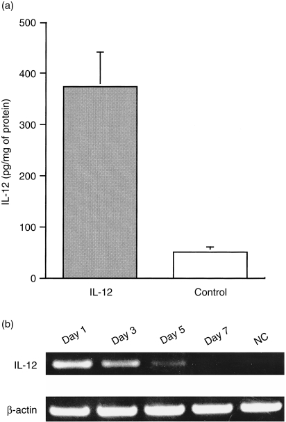
Figure 2.
Expression of the interleukin-12 (IL-12) in gene gun-treated skin tissues. Plasmid DNA (pCAGGS-IL-12 or pCAGGS) was precipitated onto 1·6-µm gold particles. Mice were shaved in the abdominal area, and abdominal skin was transfected with a 300 p.s.i. helium gas pulse by using the Helios Gene Gun. (a) Detection of IL-12 p70 protein in IL-12 gene-transfected skin tissues at 24 hr after transfection. Transfected skin tissues were removed, homogenized with extraction buffer and centrifuged at 7000 g to remove the debris. The content of IL-12 p70 in the supernatant was estimated by using enzyme-linked immunosorbent assay (ELISA), as described in the Materials and methods. (b) Detection of IL-12 mRNA in IL-12 gene-transfected skin tissues. RNA was extracted from IL-12 gene-transfected skin tissues at various time-points after treatment. cDNA products prepared from RNA were amplified by using the polymerase chain reaction (PCR). For IL-12 mRNA detection, specific primers were designed to encompass the intron sequence. The IL-12 forward primer was designed to hybridize with the sequence immediately downstream of the transcription start site of the CAG promoter and thus the IL-12 primer pair could amplify IL-12 cDNA derived from pCAGGA-IL-12, but not from endogenous IL-12. NC, sample from skin tissue transfected with the pCAGGS expression plasmid.

