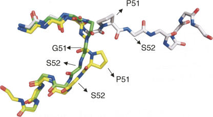
Figure 2.
Overlay of the backbone hinge region (residues 46–57) of cyanovirin, monomer and dimer, and of P51G-m4-CVN. The monomeric NMR structure of wt CV-N (PDB code 2EZM) is represented in yellow, while the domain-swapped crystal structure (PDB code 1M5M) is represented in gray, and the monomeric crystal structure of P51G-m4-CVN in green (PDB code 2RDK; this work). Only the side chain of P51 (wt) is shown. In 2EZM, S52 assumes disallowed backbone dihedral angles.

