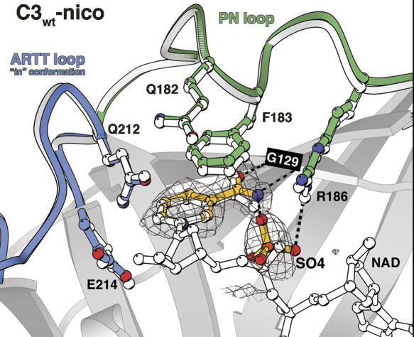
Figure 2.
The nicotinamide-binding site. Nicotinamide and sulfate ion inside the NAD binding site of the C3wt-nico structure are represented in ball-and-stick colored in yellow. The electron density grid shows an Fo–Fc omit map contoured at the 2 σ level. The ARTT and the PN loops are indicated in blue and green, respectively. Hydrogen bonds made by the nicotinamide and sulfate ion are shown by a dashed line. For comparison, the NAD and the ARTT and PN loops from the C3wt-NAD structure (1GZF) (Ménétrey et al. 2002) are superimposed and shown in white ball-and-stick representation.

