Figure 1.
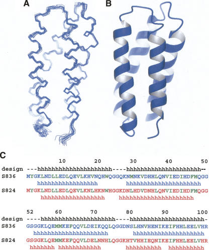
Helical backbone of protein S-836. (A) Line rendering of the 15 lowest energy structures. (B) Ribbon diagram of one representative structure. Both renderings show S-836 in the same orientation, with the N terminus in the foreground. (C) Sequence and secondary structure of protein S-836 compared with protein S-824 and with the original binary patterned design. “h” Indicates helical secondary structure. There is high sequence identity between S-836 and S-824. Differences in their primary structure occur at positions 18–35 and at positions 71–87. Helices were identified from solved structures using MOLMOL software (Koradi et al. 1996) and vary slightly from the design template. Residues that are nonpolar by binary patterning design are shown in green.

