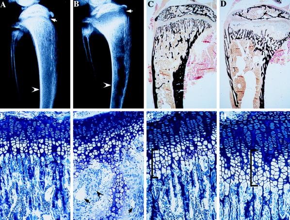
Figure 4.
Contact radiography and histology of the tibia of VDR ablated mice. (A) Contact x-ray of the tibia of a 35-day-old control mouse. (B) Contact x-ray of the tibia of a 35-day-old −/− littermate. (C) von Kossa stain of a nondemineralized section through the tibia of the 35-day-old control mouse. (D) von Kossa stain of a nondemineralized section through the tibia of a 35-day-old −/− littermate. (E) Toluidine blue section through the growth plate of the 35-day-old control mouse. (F) Toluidine blue section through the growth plate of the 35-day-old −/− littermate. (G) Toluidine blue section through the growth plate of a 15-day-old control mouse. (H) Toluidine blue section through the growth plate of a 15-day-old −/− littermate.

