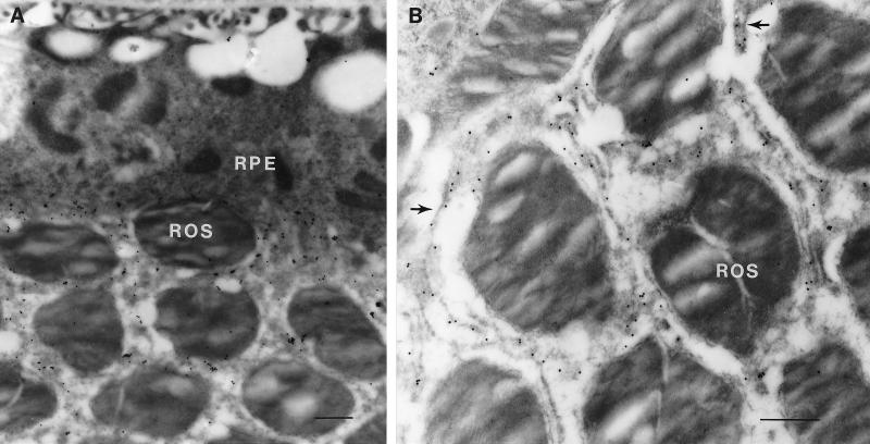
Figure 6.
Immunoelectron microscopic localization of peropsin in the mouse eye. Immunogold labeling with anti-peropsin antibodies is localized to the apical face of the RPE and to the microvilli that surround the rod outer segments (ROS). (A) At low magnification, the full thickness of the RPE is seen. Numerous large phagosomes are present within the RPE, and the infoldings at the basal face of the RPE are seen immediately adjacent to Bruch’s membrane at the top of the photomicrograph. (B) At higher magnification, individual immunolabeled microvilli (arrows) are seen between unlabeled rod outer segments. (Bars = 0.5 μm.)

