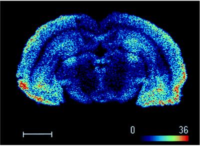
Figure 3.
Digital autoradiogram of [3H]-5-HT-moduline binding sites on rat midbrain section obtained with the β-imager. Brain sections were labeled as described in text with 1.5 nM of [3H]-5-HT-moduline and β-particles emitted were detected by β-imager. Note that binding is concentrated in regions such as cortex, central gray, hippocampus and substantia nigra. (Bar = 2.6 mm.)

