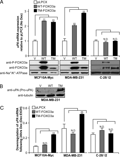
Figure 4.
uPA mRNA and protein levels after ectopic FOXO3a expression. MCF10A-Myc and MDA-MB-231 cells were stably transfected with either pLPCX, pLPCX-WT-FOXO3a, or constitutively active pLPCX-TM-FOXO3a and C28/I2 cells were transiently transfected with the same vectors. A, MCF10A-Myc, MDA-MB-231, and C-28/I2 uPA mRNA expression without Dex treatment evaluated by quantitative real-time RT-PCR. Data are shown relative to pLPCX alone after normalization to GAPDH and reported as a mean ± se of triplicate experiments. Lysates were also evaluated for FOXO3a and phospho-FOXO3a expression by Western blot. B, Lysates from MDA-MB-231 cells in the absence of Dex treatment were evaluated by Western blot to determine pro-uPA protein levels. C, MCF10A-Myc, MDA-MB-231, and C-28/I2 uPA mRNA expression with 24 h Dex (10−6 m) or vehicle (ethanol) treatment determined by quantitative real-time RT-PCR. The fold change of gene expression is shown relative to vehicle after normalization to GAPDH and reported as a mean ± se of triplicate experiments. **, P < 0.05 for FOXO3a constructs compared with control pLPCX using a two-tailed unpaired Student’s t test. ND, No significant difference compared with pLPCX; TM, TM-FOXO3a; V, pLPCX vector; WT, WT-FOXO3a.

