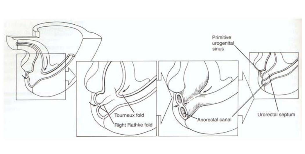
Figure 2.
Depiction of human embryonic development at weeks 5–7 of gestation. The allantois and hindgut drain into the cloaca. Classic embryology teaching suggests the cloaca is septated by the caudal and ventral growth of the Tourneux fold and the lateral to medial growth of the left and right Rathke folds. These folds coalesce, forming the urorectal septum which divides the primitive urogenital sinus from the anorectal canal. This process is controversial. Reproduced with permission41.

