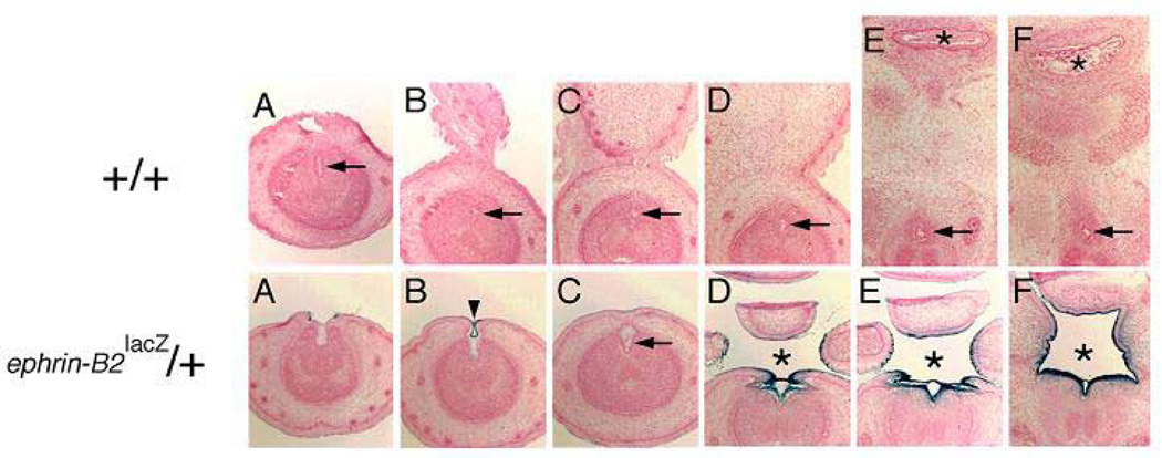
Figure 7.
Upper panel: consecutive cross sections of embryonic day 17 (E17) wild-type mouse penis demonstrating tubularized urethra (arrow) and normal septated anus (asterisk). From left to right, the sections progress from the distal penis inward into the perineum. Lower panel: consecutive cross sections of embryonic day 17 (E17) ephrin-B2lacZ/+ heterozygous mouse penis demonstrating incomplete proximal urethral tubularization and perineal closure (asterisk). In some more distal locations, urethral tubularization appeared more normal (arrow and arrowhead). The dark-blue staining indicates the high expression of ephrin-B2lacZ in the tubularizing urethral epithelium, septating epithelium of the cloaca and urogenital sinus (not shown). From left to right, the sections progress from the distal penis inward into the perineum. Reproduced with permission39.

