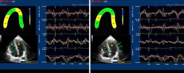
Figure 1.

2D LPSS evaluation in athletes' at rest (left side) and after HG (right side). The endocardial border of the 2D left ventricular chamber is automatically followed in time frame by frame. There is no obvious differences in strain in basal and apical segments at rest, but a significant increase after HG.
