Figure 2.
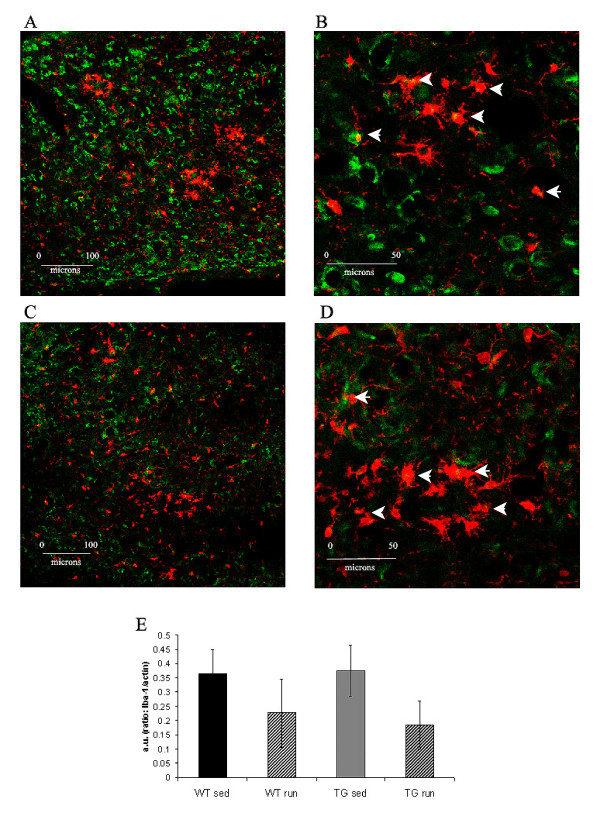
CD11b positive microglia (green immunofluorescence) in TGSED(A). Higher magnification reveals some co-labeling with microglial marker Iba-1 (red) (B, arrowheads). CD11b positive glia are present in TGRUN (C) and co-labeled with Iba-1 (red) in some cases (D, arrowheads). Overall levels of Iba-1 (normalized to actin) are not significantly different based on condition or genotype (E). High immunoreactivity for Iba-1 in WT is likely due to the advanced age of the animals used.

