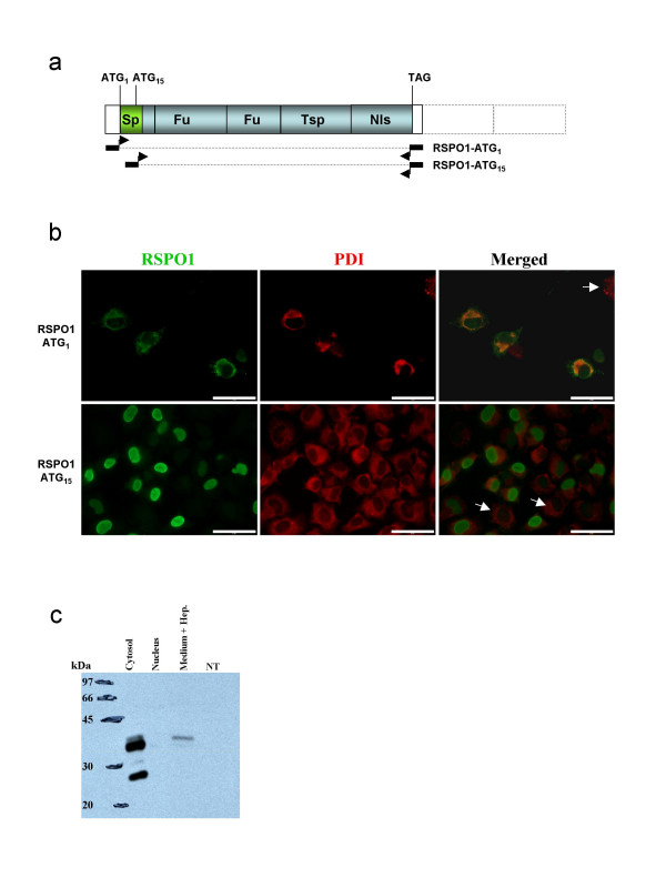
Figure 4.
Test of the RSPO1 antibody specificity by immuno-florescence studies and Western blotting. a) Schematic representation of goat RSPO1 cDNAs used in transfection experiments in COS7 cells. The longest comprise the first initiator codon (ATG1). The smallest encodes a putative protein beginning at ATG15. The black rectangles with arrows depicted the location of the primers used in order to clone both cDNAs. b) RSPO1 and Protein Disulphide Isomerase (PDI) immuno-detection in COS7 cells transfected with RSPO1-ATG1 or RSPO1-ATG15 expression vectors. Scale bars = 5 μm. c) Western blot detection of RSPO1 on total proteins extracted from the cytosolic or nuclear compartments and from the heparin-supplemented culture medium of COS7 cells transfected with RSPO1-ATG1 compared with none transfected cells (NT).

