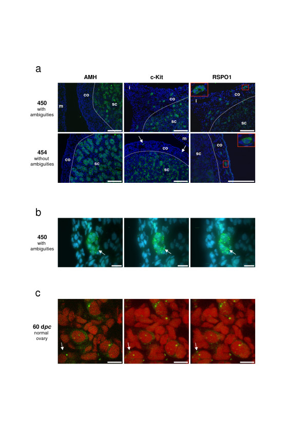
Figure 7.
RSPO1, AMH and c-Kit immuno-detection on goat sex-reversed gonads at 50 dpc. The fluorescent staining is presented with a DAPI blue (a, b) or a propidium iodide (c) nuclear-specific counterstaining. a) AMH (Sertoli-specific cell marker) and c-Kit (Leydig and germ cells marker) detections reveal the difference in testis development between XX male with ambiguities (N°450) and without ambiguity (N°454). In both cases, only the germ cells located in the cortical region (co) outside the seminiferous tubules show a RSPO1 specific staining. Note the presence of germ cell colonies in XX male with ambiguities (insert) and the presence of isolated germ cells in XX male without ambiguity (arrows for c-Kit staining and insert for RSPO1). Inserts correspond to a 3.0 enlargement of the red rectangles depicted on the same picture. m = mesonephros; i = isthmus between gonad and mesonephros; sc = sub-cortical region. Scale bars: 100 μm. b) Confocal microscopic views of a germ cell colony (10 cells) located in the tunica albuginae of the XX testis (N°450), after RSPO1 immuno-detection. Sale bars: 20 μm. c) Confocal microscopic views of isolated meiotic germ cells in the cortical area of a 60 dpc normal ovary, after RSPO1 immuno-detection. Scale bars: 10 μm. b-c) Left picture is a view of one confocal slice (0.36 μm thickness). Middle and right pictures are projected views of the different slices (n = 50) into two different angles, which allow to ascertain the punctuated staining at the cell membrane (as example the same fluorescent point is marked with an arrow in the three views).

