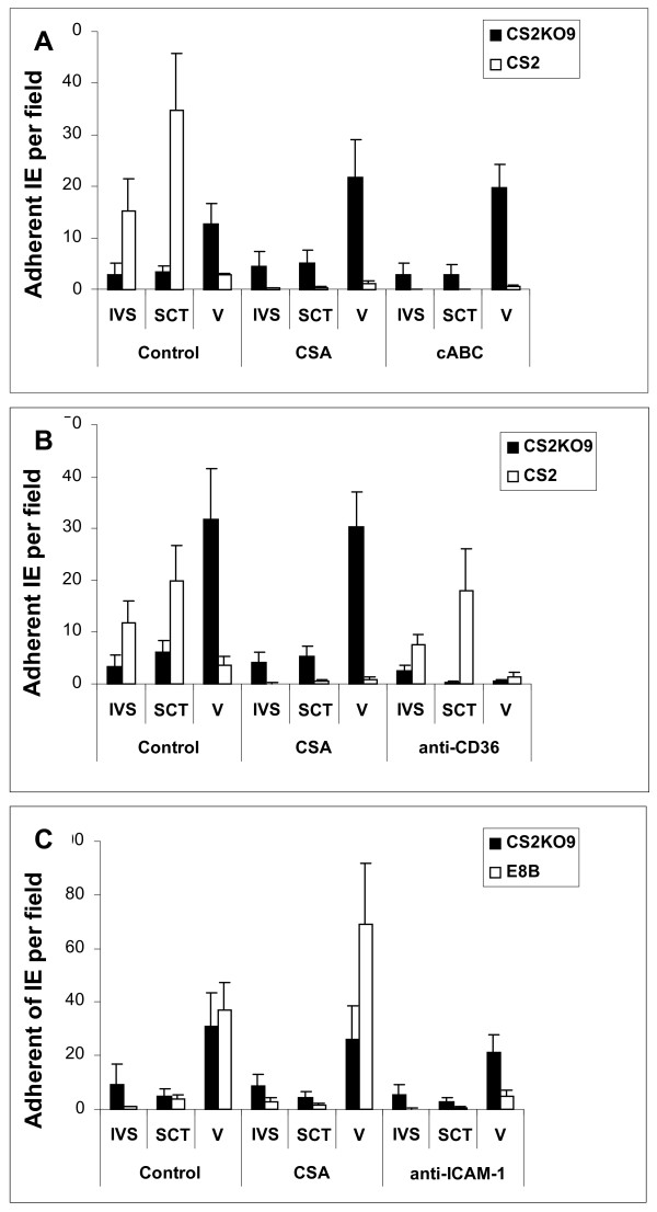
Figure 3.
Adhesion to human placental tissue cryosections. Adhesion levels of IE in the intervillous space (IVS), to syncytiotrophoblast (SCT) and over the villus (V) is shown compared to untreated (control) sections present on each slide. (a) Adhesion of CS2 (white bars) and CS2KO9 (black bars) IE in the presence of CSA (100 μg/ml) or following chondroitinase ABC pretreatment (cABC) (0.5 units/mL). (b) Adhesion of CS2 (white) and CS2KO9 (black bars) IE in the presence of CSA or following anti-CD36 pre-treatment (0.5 μg/ml). (c) Adhesion of E8B (white) and CS2KO9 (black) IE in presence of CSA or following pretreatment with anti-ICAM-1 (10 μg/ml). Adhesion was counted at 40× magnification and expressed as average IE bound per field (+SEM) for at least 3 independent experiments.

