Fig. 3.
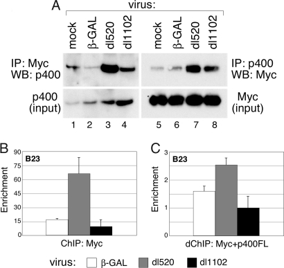
E1A expression promotes the formation of Myc–p400 complexes. (A) U2OS cells were transiently transfected with HA-tagged Myc and FLAG-tagged p400 for 48 h. Transfected cells were infected for 6 h with β-GAL, dl520, or dl1102 adenovirus. Coimmunoprecitation was performed as described (4) by using anti-Myc (N262) or anti-FLAG (M2) antibodies, and Myc and p400 were detected by WB. (B) U2OS cells were transfected with FLAG-p400 or control DNA for 48 h and then infected with β-GAL, dl520, or dl1102 adenovirus for 6 h. ChIP analysis was performed by using the anti-Myc antibody N262, and coprecipitating DNAs corresponding to the B23 E-box were detected by quantitative PCR. Enrichment is calculated relative to a non-Myc binding sequence. (C) Protein–DNA complexes recovered in B were eluted and subject to a second round of ChIP with anti-FLAG antibodies to recover p400-containing chromatin. Levels of coprecipitating DNA from the B23 E-box were determined by quantitative PCR. To control for nonspecific background, signal in the re-ChIP was normalized to that from an identical, parallel, experiment from cells not expressing FLAG-tagged p400.

