Fig. 1.
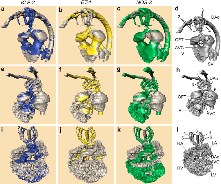
Computerized reconstructions after serial histological sections of embryos represent three subsequent stages of the developing chick heart. First row (a–d), left lateral view of Hamburger Hamilton (HH) stage18. Second row (e–h), left ventrolateral view HH24. Third row (i–l), ventral view HH27. The cardiac and vascular compartments are annotated in d, h and l, respectively. Note the disappearance of the second pharyngeal arch artery PAA (compare d, h) and the emergence of the sixth one in h. The asterisk denotes the inner curvature of the looping heart that becomes tighter during development. The outflow tract (OFT) is connected to the pharyngeal arch arteries (PAA). The OFT is located right sided in HH18, subsequently it becomes wedged between both atria and centrally localized in the cardiac contour in HH27. The stained parts in the columns represent the expression of the respective shear stress-induced genes, KLF2 (blue), ET1 (yellow) and NOS3 (green). Note that KLF2 is restricted to the narrow zones (AVC, OFT, PAA 2, 3, 4), whereas ET1 is preferentially localized in the wider segments. NOS3 is mainly absent from the wider atrial segments. Abbreviations: 2/3/4/6th 2nd, 3rd, 4th and 6th pharyngeal arch arteries, * inner curvature of the heart, A primitive Atrium, AVC Atrioventricular canal, DAo Dorsal aorta, LA left atrium, LV left ventricle, OFT Outflow tract, RA right atrium, RV right ventricle, SV sinus venosus, V primitive ventricle. Figure modified after [12
]

