
Figure 3.
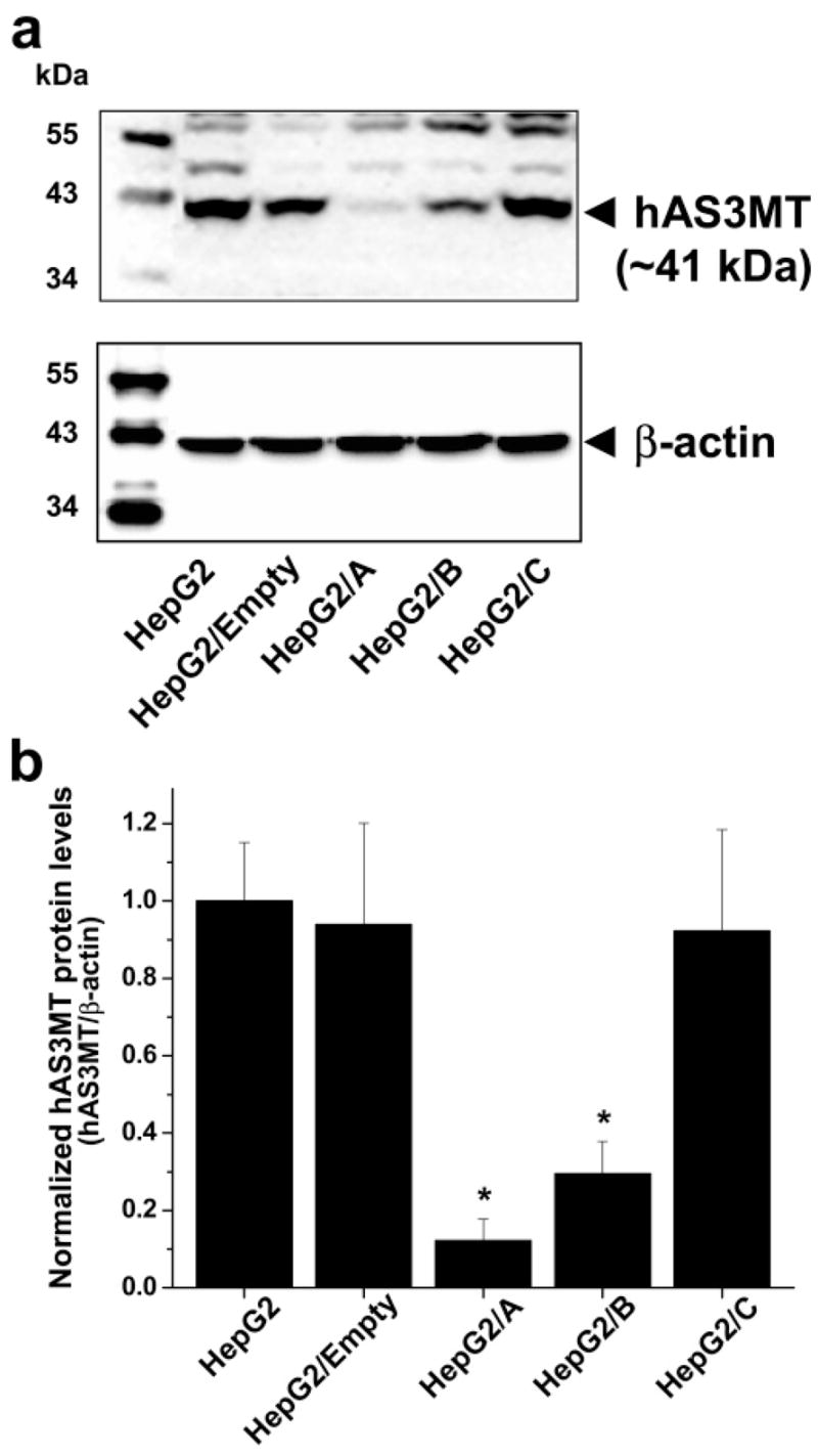
hAS3MT protein levels in parental and clonal HepG2 cells. (a) Immunoblot images of hAS3MT and β-actin. A representative image from one of three independent cultures is shown. (b) hAS3MT protein levels normalized for β-actin contents. Mean and SD values for the three independent cultures are shown. *The mean value is significantly different (p < 0.05) from that in parental HepG2 cells.