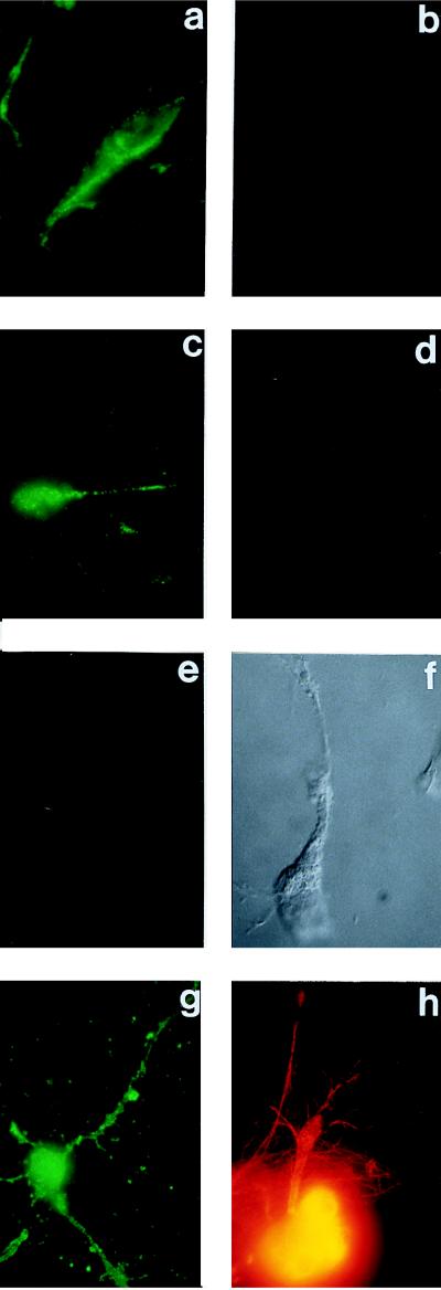
Figure 3.
Immunofluorescence microscopic labeling with either live, or fixed and permeabilized, untransfected NT2N neuronal cells, using as the primary antibody reagents either the anti-PS-1 antibodies of Fig. 1A, or anti-tubulin antibodies, or both in double-labeling experiments on the same cells. In these latter experiments, the anti-PS-2 labeling used a fluoresceinated secondary antibody, and the anti-tubulin labeling a rhodamine-tagged secondary antibody. (a and b) Live cells, double labeling with anti-PS-1 and anti-tubulin, respectively. (c and d) Another example of a and b, respectively. (e) Live cell, single labeling with anti-PS-1 in the presence of an excess of the soluble specific PS-1–MAP oligopeptide complex. (f) Nomarski image of e. (g) Fixed and permeabilized cell, single labeling with anti-PS-1. (h) Fixed and permeabilized cell, single labeling with anti-tubulin.

