
Fig. 2.
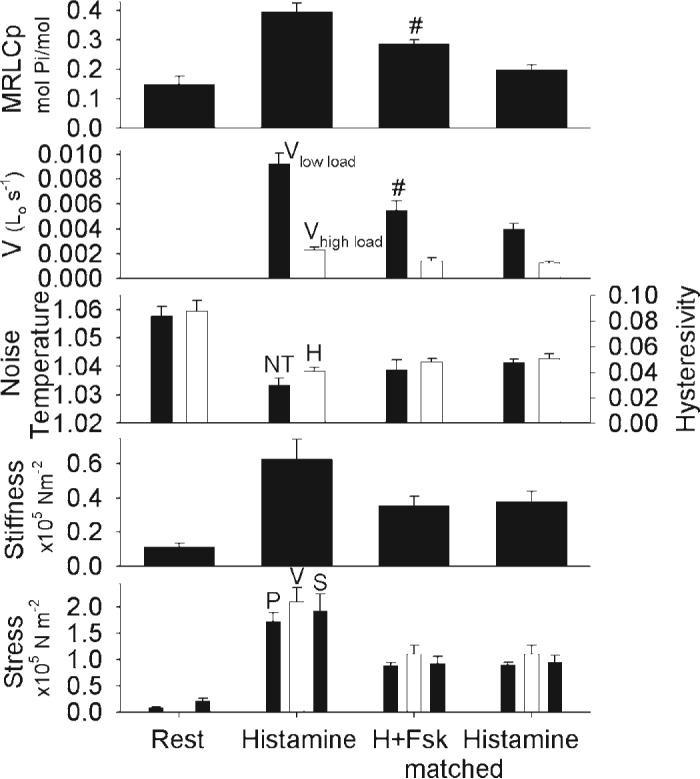
Mean biochemical and mechanical characteristics in swine carotid artery tissues with and without force suppression. Tissues were either unstimulated (column 1), maximally stimulated with 10 μM histamine for 25− 40 min (column 2), stimulated with 10 μM histamine and relaxed with forskolin to ∼50% of maximal force (varying times and forskolin concentrations, column 3, with force suppression), or stimulated with various concentrations of histamine to the same ∼50% of maximal force (column 4, without force suppression). The panels shown mean ± 1 SE for crossbridge phosphorylation [phosphorylated myosin regulatory light chain (MRLCp), panel 1], shortening velocity at low 17% load (panel 2, filled bars labeled “Vlow load”), shortening velocity at high 48% load (panel 2, open bars labeled “Vhigh load”), noise temperature (panel 3, filled bars), hysteresivity (panel 3, open bars), stiffness (panel 4), and stress [panel 5, the filled bars on left (labeled “P”) are the stress values from the MRLCp experiments and the open bars (labeled “V”) are the stress from the velocity experiments; filled bars on right (labeled “S”) are from the stiffness experiments]. Data are presented as means ± 1 SE with n = 5−6 experiments. #Significant difference when comparing tissues with and without force suppression, i.e., column 3 vs. column 4. Force suppression was associated with significantly higher crossbridge phosphorylation and velocity at low loads when compared to tissues at matched force without force suppression. Velocity at higher load, noise temperature, hysteresivity, and stiffness did not significantly differ with and without force suppression.