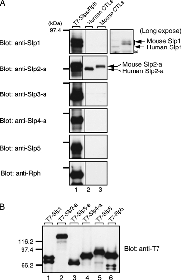
Figure 1. Expression of Slp1 and Slp2-a in CTL.

A) Western blots showing mouse and human CTL lysates probed for expression of Slp1-5 and rabphilin (Rph). Lanes show control lysates of T7-tagged Slp1-5 or Rph (lane 1) and lysates from human (lane 2) and mouse CTLs (lane 3). Blots are probed with antibodies against Slp1-5 and Rph as described in Imai et al. (37). The marker on the left shows the relative migration of the 97.4 kDa molecular weight marker. A longer exposure of Slp1 is shown on the right. Note that the Slp1 and Slp2-a proteins, but not the other Slp proteins, were detected in human and mouse CTLs (indicated by arrows). Asterisk corresponds to non-specific bands. B) Recombinant T7-tagged Slp1-5 and Rph were used as positive controls in A. Similar amounts of the T7-tagged proteins from COS-7 cell lysates were loaded into each lane and probed with anti-T7 tag antibody as described previously (37). Relative migration of molecular weight markers (in kiloDalton) is shown on the left.
